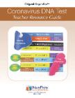
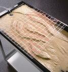
Coronavirus DNA Test 3-D Model Kit-10 Student Model Template Sets and Teacher Guide, Vinyl Pouch, Assemble and use 3-D models to visualize and investigate key science structures, Perfect for use in classroom, lab or at home, Satisfies NGSS Standards, Grade: 6-10, Material: Paper, LxW: 12X19in
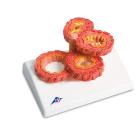
Model Copd, is a chronic lung disease with severe narrowing of the airways, Dimensions: 15x11x8 cm, Even administering medication cannot fully cure this narrowing, normal condition, abnormal mucous secretion, thickening of the bronchial mucosa, permanent damage
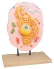
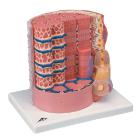
Three dimensional model showing electron microscopic structure. Organs like nucleus, Nucleolus, endoplasmic reticulum, mitochondria, ribosomes respectively polysomes and golgi apparatus. Showing centrioles, lysosomes and vacuoles.
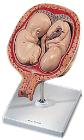
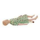
Model, 5th Month Twin Fetuses - Normal Position, shown in a natural position within the uterus, They can be removed for a closer inspection of the uterus, this high quality model is a great tool for studying the anatomy of human development, weight: 0.66 kg
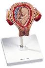
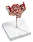
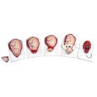
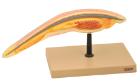
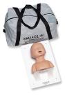
Model, Embryo, 3 month, The embryo model shows an embryo at the stage of the first month of pregnancy, Ideal for medical training and as an educational tool for pregnant women, Important anatomical structures are labeled
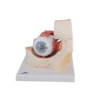
Model, Embryo, 1 month, The embryo model shows an embryo at the stage of the first month of pregnancy, Ideal for medical training and as an educational tool for pregnant women, Important anatomical structures are labeled, weight: 0.33 kg
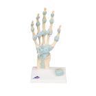
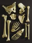
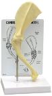
Model Hand Skeleton, with Ligaments and Carpal Tunnel, Dimensions: 30 x 14 x 10 cm, shows the anatomical detail of the ligaments and tendons found in the hand, wrist, and lower forearm, The interosseous membrane between the radius and ulna with the bones
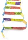
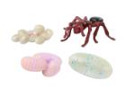
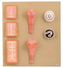
Ant Life Cycle Stages, Children can see how ants change as they grow with Insect Lore’s Ant Life Cycle Stages. These oversized, anatomically correct figures have been accurately painted and sculpted to show the four stages of ant development: eggs, larva, pupa and adult.
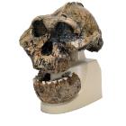
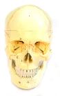
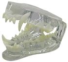
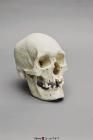
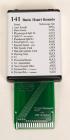
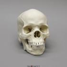
Each part on the skull is numbered. Jaw is spring loaded. Skull cap is removable for further interior inspection. Included with the model is a sheet with each numbers name for identification.
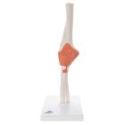
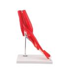
Model Elbow Joint 8 Parts, great tool for student and patient education, part of a high quality series of muscle models, and has been manufactured to replicate the anatomy of the human elbow joint in detail, colors used, Weight: 1.74 kg, Dimensions : 25 x 41 x 25 cm
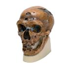
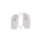
Human Male Polynesian Skull, sagittal keel, parietal bossing, the broad, prominent basiooccipital, and the rocker jaw, rocker jaw is curved along the inferior surface of the mandible, 2-part skull (separate cranium & jaw), Size: 20.7x14.2x19.6cm
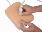
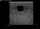
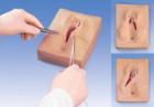
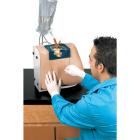
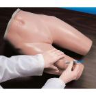
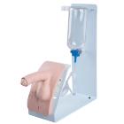
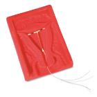
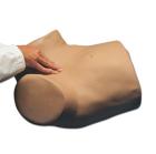
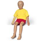
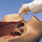
Catheterization Simulator Basic, With the male P93, Male catheterization with realistic resistance, 3 levels of adjustable narrowing of urethra, Soft and movable foreskin, Liquid outflow if catheterization is successfully carried out, Easy to clean
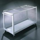
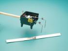
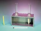
Convection of Gases Apparatus, consists of a metal box (22x10x11cm) with clear acrylic front panel, two glass chimneys, a candle and a cotton filled wick as the smoke source, Includes activity guide
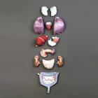
Soft Organ Set, Stainless-steel fittings, 100% non-breakable plastic bones repli-cast from those of a select male specimen, Detachable arms and legs, Detachable calvaria for access to the neuro-cranium, Hinged mandibular flap which opens to expose roots of teeth in their sockets
Model Human Male African-American Skull, a depressed nasal root, obtuse nasal angle, short anterior nasal spine, bilateral gutter at lower part of nasal aperture, rectangular-shaped palate & blade-like incisor in upper jaw, Size: 20.9x13.7x20cm
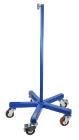
Support base for skeleton, features a heavy (30 pound) cast iron base with 5 machine grade casters (2 with locks) and a sturdy threaded pole, this skeleton stand will pass the test of time and make the transport of y skeleton easy. Inner diameter of stand is 12.5mm.