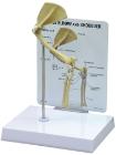
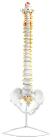
Somso Special Natural size, in SOMSO-Plast*. Nerve endings, filum terminale and cauda equina of the spinal cord (also in cross-section) are shown. Separates into 2 parts. On a stand with green base.
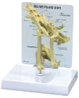
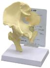
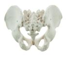
It Illustrate the morphological distinctions between male and female pelvic structures. Each pelvis includes the left and right innominates with pubic symphysis, 4th and 5th lumbar vertebrae with intervertebral discs, the sacrum and coccyx.
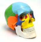
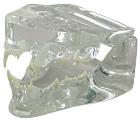
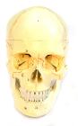
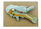
Each part on the skull is numbered. Jaw is spring loaded. Skull cap is removable for further interior inspection. Included with the model is a sheet with each numbers name for identification.
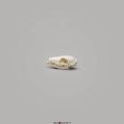
Amoeba proteus, enlarged approximately 1000 times. In a small pseudopodium which can be opened up showing the structure after electron microscopic magnification. On a base with explanatory notes. Separates into 2 parts
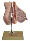
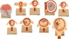
Set of nine models, showing the following stages. 1. Embryo 6 days old 2. 1st month of gestation. 3. Uterus with embryo in 3rd month of gestation.4. Uterus with fetus, in 4th month. 5. Uterus with fetus, placenta and umbilical cord.6. 5th month. 7. 7th month pregnancy.