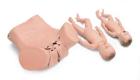
Simulator Prostate Exam 4 Stages, Trainer is used in medical simulation and clinical training for the demonstration and practice of digital rectal examinations (DRE) index finger is inserted into the rectum, facilitating palpation of the rear part of the prostate
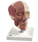
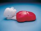
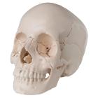
Skull model with face musculature, The face and mastication muscles are illustrated on the right half of this skull model. The face musculature can easily and precisely be differentiated from the mastication musculature by using two colours
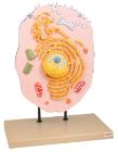
Three dimensional model showing electron microscopic structure. Organs like nucleus, Nucleolus, endoplasmic reticulum, mitochondria, ribosomes respectively polysomes and golgi apparatus. Showing centrioles, lysosomes and vacuoles.
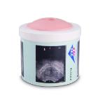
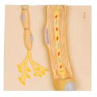
Model Schwann Cells Of The PNS, depicts a Schwann cell with sectioned core, depicts the schwann cell in colorful anatomical detail, great addition to any lesson on the human nervous system, Weight: 0.4 kg, Dimensions : 12.2 x 11.7 x 3.2 cm
Model Physiology Of Nerves Series, displays the basic structures of the human nervous system, Each of the five sections of the nerve model shows a plastic colored relief model of the main synapse variations, Weight: 4.379 kg, Dimensions : 68 x 51 x 3 cm