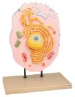
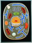
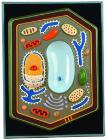
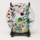
Three dimensional model showing electron microscopic structure. Organs like nucleus, Nucleolus, endoplasmic reticulum, mitochondria, ribosomes respectively polysomes and golgi apparatus. Showing centrioles, lysosomes and vacuoles.
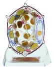
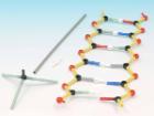
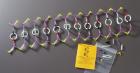
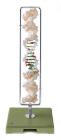
Meiosis Model, Dimensions: 60 x 40 x 6 cm, Advantages: Chromosomes coloured according to modified AZAN staining colour, Cell components are colour-coded in accordance with educational aspects, Attaching magnets on the rear, Storage system, Enlarged 10, 000 times
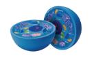
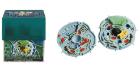
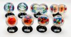
Science BioSigns Animal Cell Model, Hands-on interactive 14-piece model teaches about cell membranes and mitochondria, Provides a magnified and cross-sectioned detailing of an Animal Cell, Includes Guide
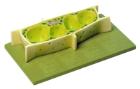
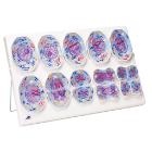
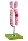
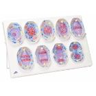
Mitosis Model, Dimensions: approx. 60x40x6 cm3, Advantages: Chromosomes coloured according to modified AZAN staining colours, Cell components are colour-coded in accordance with educational aspects, Attaching magnets on the rear, Storage system, Enlarged 10, 000 times