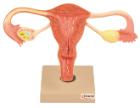
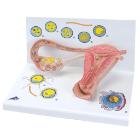
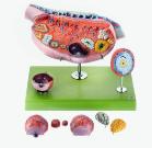
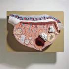
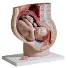
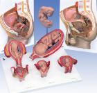
Model Stages Of Fertilisation, Dimensions: 35x21x20 cm, begin the growth into an embryo, The various stages are shown in larger-than-life model form in an ovary, fallopian tube and womb. An even more enlarged illustration of each is also printed on the base
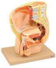
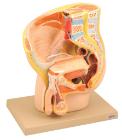
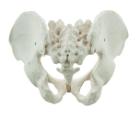
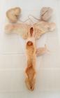
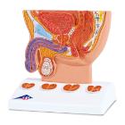
It Illustrate the morphological distinctions between male and female pelvic structures. Each pelvis includes the left and right innominates with pubic symphysis, 4th and 5th lumbar vertebrae with intervertebral discs, the sacrum and coccyx.
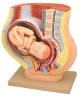
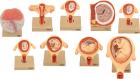
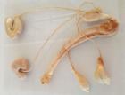
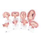
Set of nine models, showing the following stages. 1. Embryo 6 days old 2. 1st month of gestation. 3. Uterus with embryo in 3rd month of gestation.4. Uterus with fetus, in 4th month. 5. Uterus with fetus, placenta and umbilical cord.6. 5th month. 7. 7th month pregnancy.
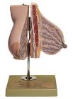
Model of the Ovary, Enlarged approximately 10 times, in SOMSO-Plast* Horizontal section parallel to the mesovarian margin with presentation of the follicles in different maturation phases, corpus rubrum, luteum and albicans, Separates into 13 parts. Mounted